Welcome to the Längst Lab - Applied Epigenetics
We are part of the Institute for Biochemistry, Genetics and Microbiology and member of the Regensburg Center for Biochemistry (RCB) at the University of Regensburg.
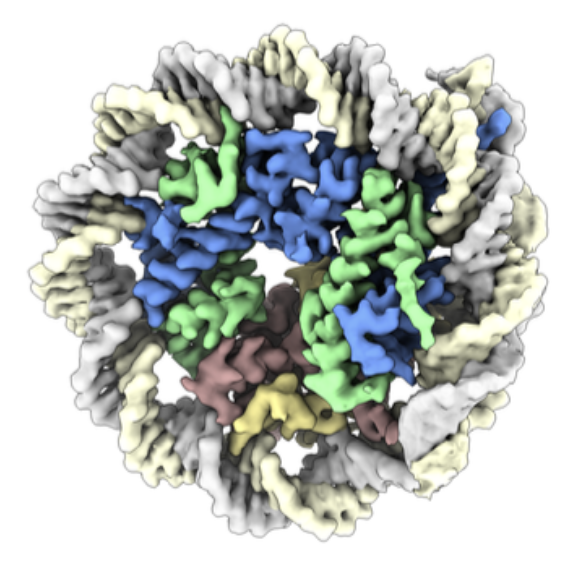
The Längst Group investigates the mechanisms and dynamics of packaging the eukaryotic and viral genetic material into chromatin and the latter's function in disease processes.
This research includes human chromatin, the genetic packaging of Adenovirus and Coronavirus, and chromatin structure dynamics in the Malaria-causing parasite Plasmodium falciparum.
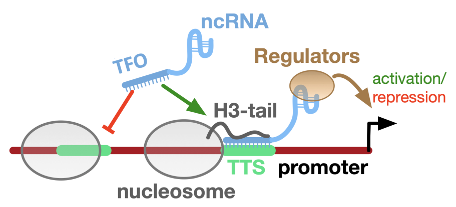
While our fundamental research focuses on chromatin remodelling complexes and non-coding RNAs in nuclear architecture, nucleosome positioning and gene regulation, our applied research relates to inhibiting the dynamics and packaging of the genetic material to identify new drug targets and block cancer, pathogen and viral growth.
News
July 2026
Holzinger et al., Plos Computational Biology (2026) link to article (PMID: 42507729)
Deciphering chromatin architecture and dynamics in Plasmodium falciparum using the nucDetective pipeline
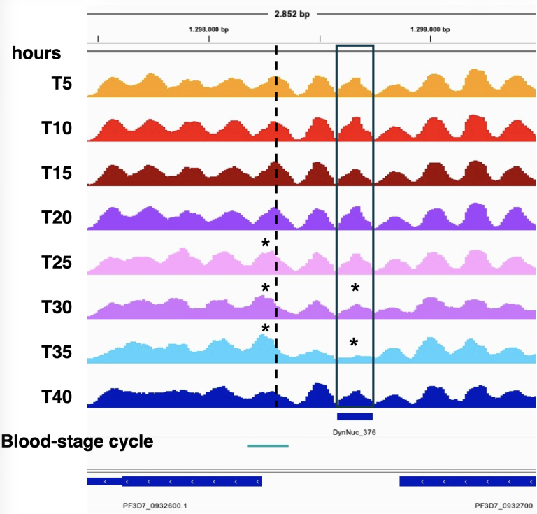
Contrary to the current view that this parasite has irregular chromatin, we discovered regular phased nucleosome arrays downstream of transcription start sites. Along with the established +1 nucleosome and a nucleosome-depleted region upstream, these nucleosome arrays form a promoter structure similar to that of other eukaryotes. Nucleosome spacing varied between developmental stages, and nucleosomal rearrangements occurred in intergenic regulatory regions. These dynamic nucleosomes were associated with histone modifications, histone variants, DNA accessibility, gene expression, and transcription factor binding sites. Our findings reveal an underappreciated level of specific and dynamic chromatin organisation in the malaria parasite that aids understanding of developmental processes and helps identify therapeutic targets.
Understanding how DNA is packaged inside cells is crucial for studying gene regulation during development. In eukaryotes, DNA wraps around protein spools to form nucleosomes. The precise positioning of these nucleosomes along the genome serves as a key layer of gene regulation. We developed nucDetective, a computational pipeline that maps nucleosomes across the genome and compares their organisation between samples. It measures nucleosome positioning, spacing, occupancy, fuzziness, and array regularity. We applied nucDetective to the malaria parasite Plasmodium falciparum and analysed nucleosome organisation throughout its developmental stages inside human red blood cells.
June 2026
Adi Danieli-Mackay et al., Cell Reports (2026), accepted
Targeting addiction to HMGB2-driven transcriptional programs in pancreatic cancer
Pancreatic cancer remains at a stagnant 5-year survival of <13% attributed to the high heterogeneity and plasticity of these tumors. To circumvent this, we focus on the abundant nuclear protein HMGB2. HMGB2 depletion is key for establishing replicative senescence in normal cells, but it is significantly overexpressed across multiple cancer types. Here, we combine single-cell and spatial genomics with patient-derived organoids and tumor samples to show how increased HMGB2 availability represents a transcriptional addiction fueling cell cycle progression and growth. We repurpose a small molecule inhibitor targeting HMGB2 to interfere with its binding to DNA, restrict chromatin accessibility at promoters, and constrain tumor growth both in vitro and in vivo. 3D chromatin interactions involving HMGB2-bound enhancers and promoters also collapse upon drug treatment. Thus, pharmacological HMGB2 targeting represents a universal strategy for managing cancer progression irrespective of its genetic or molecular characteristics.
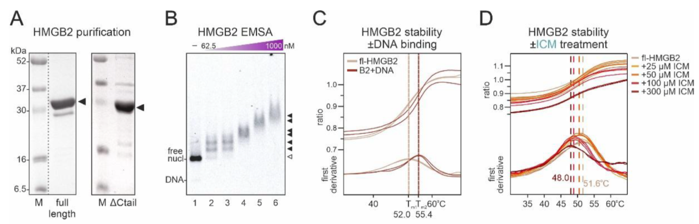
Characterization of HMGB2 association with chromatin. (A) SDS-PAGE and Coomassie blue staining of purified full length (left) and ∆Ctail HMGB2 (right) with protein marker (M) sizes indicated. (B) Electromobility shift assay (EMSA) using Cy5-tagged nucleosomes with a DNA linker (600 fM) with increasing concentrations of HMGB2 (lanes 2-6). The migration positions of free DNA, nucleosome (open triangle) and higher-order nucleosome::HMGB2 complexes (closed triangles) are indicated. (C) Stability of 15 µM full-length HMGB2 assessed by nanoDSF, where intrinsic tryptophan fluorescence was measured at 330 and 350 nm over a temperature gradient from 20°C to 95°C in the absence or presence of DNA. Deduced T m values are shown at the bottom of each graph. (D) As in panel C, but in the presence of increasing concentrations of ICM.
Biophysics Facility News
March 2026
Harald Wodrich and Gernot Längst BioSpektrum
(2026) link to article
Dynamische Veränderung der Adenovirus-DNA-
Verpackung führt zur Aktivierung
Special Genetik&Epigentik
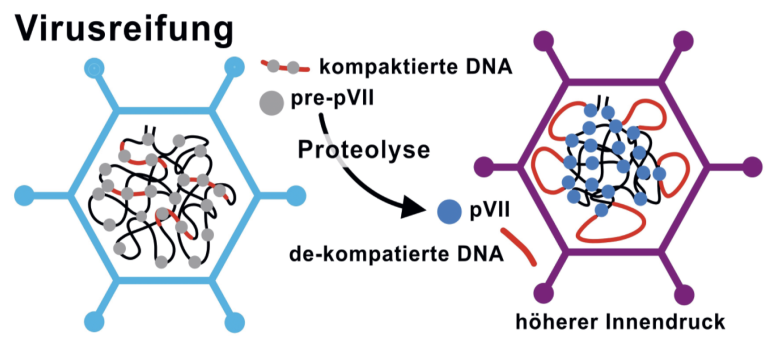
Human adenoviruses are tools for gene therapy and vaccination. The viral DNA genome is compacted by protein VII inside the capsid. We show that decompaction of the viral genome and acquisition of host nucleosomes at early promoters enables efficient viral transcriptional activation. Decompaction starts upon maturation, directed by a DNA sequence code, pre-arranging the genome for capsid release and rapid transcriptional activation, highlighting an exceptional example of evolutionary adaptation.
Teaching
We are organising basic and advanced teaching activities. Lectures, seminars, and practical courses are offered for medical students, BSc and MSc students in biology, biochemistry, and molecular medicine.
Graduate School
Gernot Längst is the managing director of the International Graduate School Regensburg (RIGeL). All PhD students of the Faculty NWFIII are members of RIGeL.
Biophysics
Our lab hosts a Biophysics Facility to characterize and quantify all kinds of molecular interactions. The facility is accessible to all UR groups.
Bioinformatics
The Bioinformatic Core Service (GACT) is associated with our group. It is supporting our faculty with advanced bioinformatics regarding high-throughput sequencing analysis.
We are developing Software tools to analyze chromatin structure and to predict sequence specific formation of RNA-DNA triple helices.